Stratasys Unveils 3D Printed Dental Anatomical Model Preset
⚓ p3d 📅 2026-02-26 👤 surdeus 👁️ 11

Stratasys has been working on digital anatomy for a number of years now. The company wants to make models that look and feel like the real thing. For doctors and dentists, this could mean more accessible testing. What’s more, applications in specific pathology, super rare cases, and lots of different parts can be made relatively inexpensively. This means that students could more easily get access to the rare, complex, and difficult.
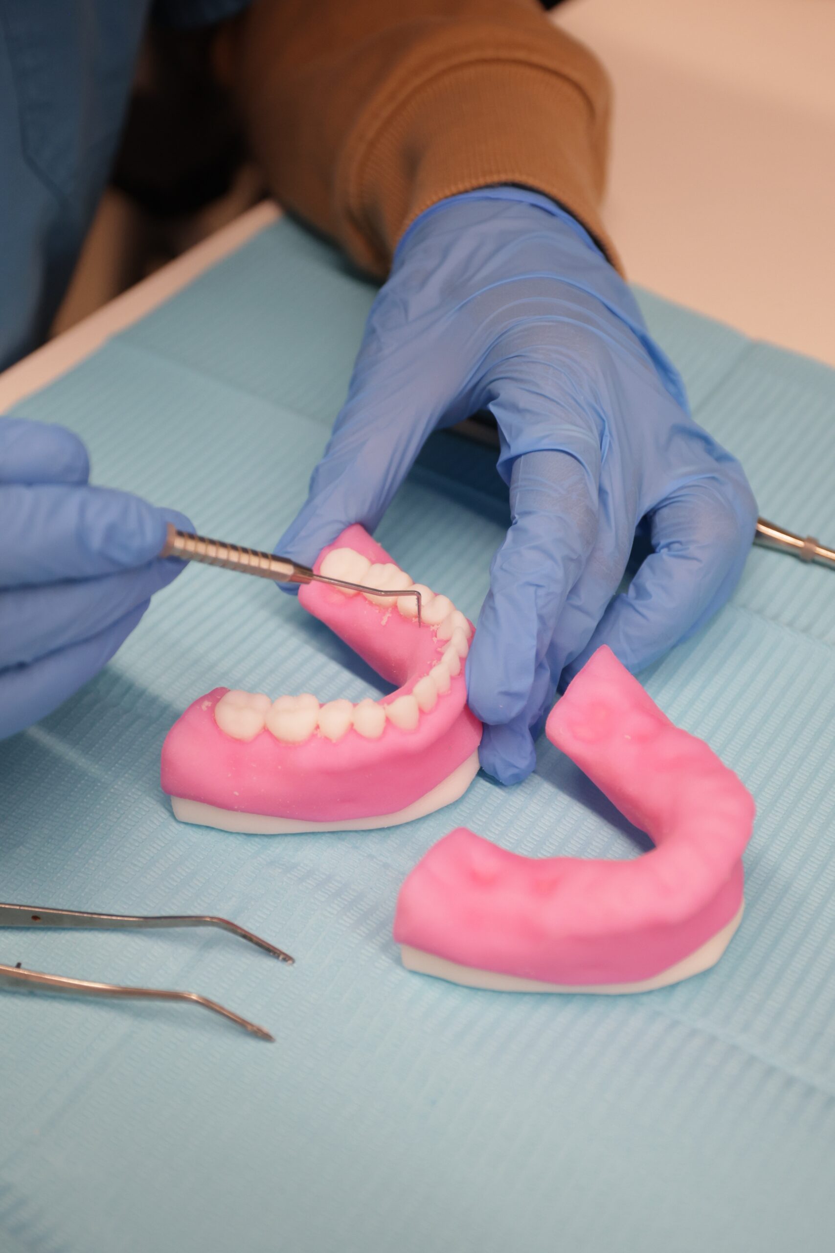
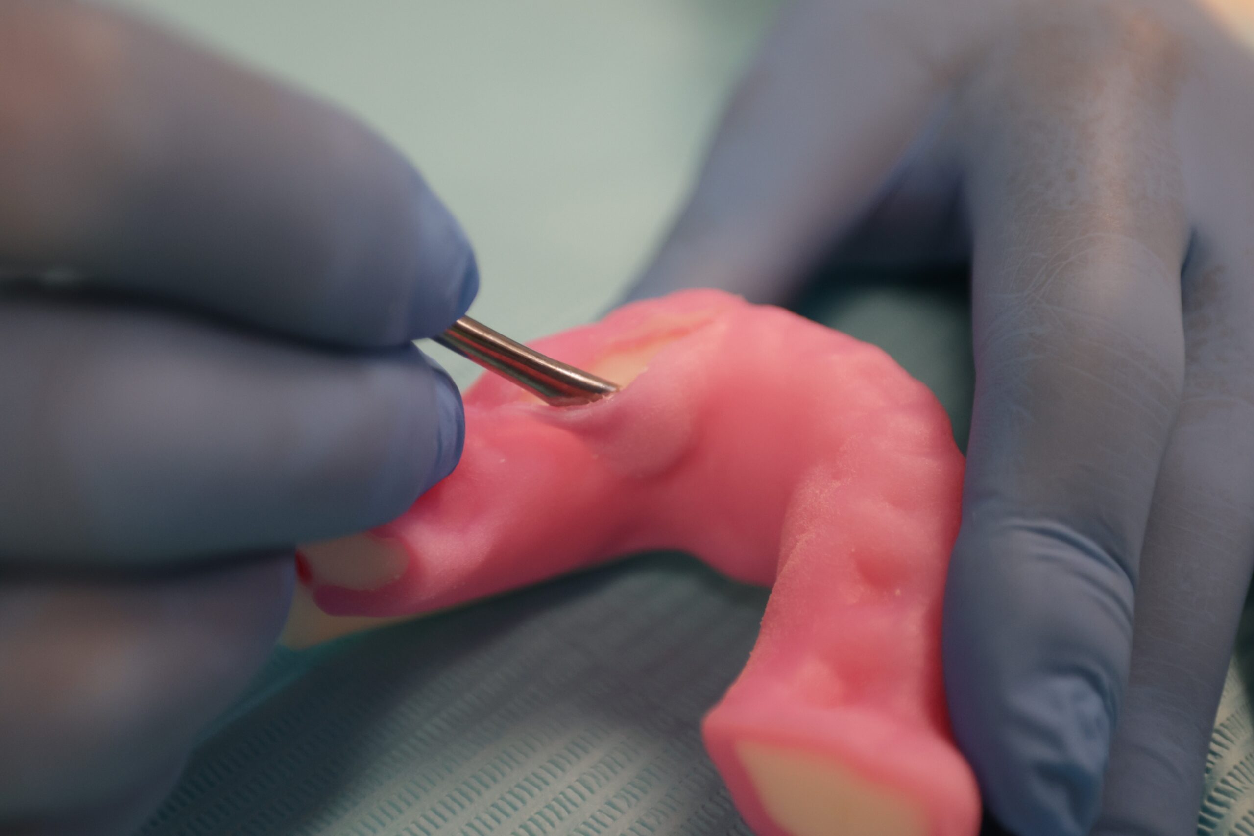
Digital anatomy replaces parts from animals, cadavers, or conventionally manufactured pieces. Cost is lower, overall training is less icky because you’re not experimenting on a body or dog mandible, and structures could closely resemble those that you will be dealing with in your practice. The company has been working for a few years on getting not only the look right through its PolyJet process, but also to get the feel just right. For your fingers, probes, and scalers, the tissue should feel just like the real thing. Ideally, Stratasys can give you a feel of that pathology so you’ll exactly know it for when you get to see and touch it in real people. Now, a 3D printed preset for dental will aid in this process.
VP Medical at Stratasys Erez Ben Zvi said,
“With this preset for dental anatomical models, we are entering a new segment of digital dental education and clinical simulation, helping customers move beyond traditional training methods toward more standardized, technology-driven learning environments. By combining anatomical realism with repeatability and customization, we’re enabling educators, clinicians, and device manufacturers to prepare for real-world procedures with greater confidence and consistency.”
The preset can be used to make a wide range of anatomical structures. The company says that, “The models replicate the biomechanical behavior of bone, teeth, nerves, and soft tissue, providing realistic haptic feedback for drilling, cutting, suturing, and implant placement.”
Trainers can use Cone Beam Computed Tomography (CBCT) scan information to make their own specific models. The CBCT scanner is that dental CT scanner that moves around your head while you bite down on a plastic bit. This kind of scan gives a 3D view of structures in your mouth, providing doctors with a far clearer picture of the actual situation in your head. CBCTs are especially useful in implants, root canals, and braces.
Stratasys suggests that these models could be used for complex cases such as “atrophic jaws, sinus lifts, and bone grafting procedures,” as well as the more quotidian “tooth extractions, implant placement, periodontal surgery, endodontic surgery, and sinus augmentation.”
Better cases, and more of them, can be a real driver for the adoption of this preset. The idea that at any given time, all your students can practice on the same complex case, for example, and then collectively share their experience, is very compelling. You could see a huge variety of shapes and pathologies as well. I’d personally work a lot better with a 3D printed model than a body or part of an animal. I also like that teachers can turn to cases they themselves have experienced as a learning tool. Maybe one type of root canal should not be done at all, or is so tricky that the patient should come back another time. This is the kind of situation that can be found in scans easily and then given to students.
I don’t know if dentists feel that these models are accurate for them. Does it really feel like the human mouth does? Is the pass of your mirror over the gums the same? But if it comes close, then this can be a viable, less expensive training solution for dentists the world over.
🏷️ p3d_feed