3D Printing News Briefs, July 26, 2025: Corrosion Resistance, 3D Printed Stents, & More
⚓ p3d 📅 2025-07-26 👤 surdeus 👁️ 32

This weekend’s 3D Printing News Briefs are all about research! We’ll start with what’s been called the first comparison of corrosion resistance in 3D printed magnesium and zinc alloys. Then, scientists at the recent European Society for Organ Transplantation Congress reported a major step forward in diabetes research, and researchers in Korea developed and validated a 3D printed skin imitation layer for radiation dosing. Finally, Lithuanian researchers are 3D printing small stents using two-photon polymerization.
Study Compares Corrosion Resistance in 3D Printed Mg and Zn Bioalloys
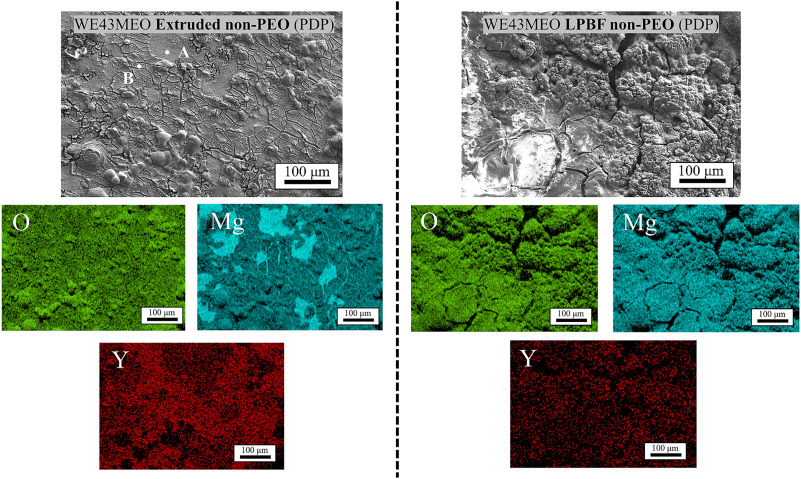
SEM images and EDS maps of the WE43MEO corroded surfaces after the PDP tests. Left side of the panel corresponds to the extruded sample and right side to the LPBF sample.
Researchers from the IMDEA Materials Institute, the Helmholtz-Zentrum Hereon Institute of Surface Science, and Meotec GmbH conducted what they call the first comparison of corrosion resistance in bioabsorbable WE43 magnesium and Zn1Mg zinc alloys produced with material extrusion and laser powder bed fusion (LPBF) 3D printing. Their study is also said to be the first to use electrochemical testing in a buffered saline solution to compare how the degradation of these biodegradable metals is affected by their production methods. The experimental portion happened under the Horizon Europe BIOMET4D project. The team found that the LPBF printed samples corroded much faster than the extruded ones: in the WE43 samples, yttrium oxide particles weakened the corrosion layer’s protective effect, and with the Zn1Mg, an increased volume of eutectic phases sped up microgalvanic degradation. A plasma electrolytic oxidation (PEO) surface treatment was applied to all samples to form a protective oxide layer and improve their corrosion resistance. The results could lay the groundwork for safer, long-lasting biodegradable implants.
“However, the WE43MEO LPBF specimens showed high corrosion rates despite PEO treatment, which was linked to heterogeneities in oxide layer thickness,” explained first author Guillermo Domínguez. “In contrast, PEO treatment had the opposite effect on Zn1Mg samples, where LPBF specimens demonstrated greater corrosion resistance than the extruded ones.”
ESOT Researchers 3D Print Insulin-Producing Pancreas Cells

A human pancreatic islet visualised using double immunostaining with glucagon antibody (red) and insulin antibody (blue). Credit: Afferent (CC BY-SA 3.0)
At the European Society for Organ Transplantation (ESOT) Congress 2025, a team of scientists announced an exciting breakthrough that could result in more effective, and less invasive, type 1 diabetes (T1D) treatment. They were able to successfully 3D print human islets (insulin-producing cell clusters in the pancreas) using a novel bioink made from decellularized human pancreatic tissue and alginate. Traditionally, islet transplants are infused into the liver, which often results in major loss of cells and only limited success. These researchers designed the 3D printed islets to be implanted just under the skin, which is a much less invasive procedure. A slow print speed of 20 mm/minute and low pressure (30 kPa) kept the islets safe during printing and helped them keep their natural shape. This resulted in high-density, durable islet structures that remained alive and functional in laboratory tests for up to three weeks. A porous architecture enhanced the flow of oxygen and nutrients to the embedded islets, which promoted vascularization and helped maintain cell health. Finally, the 3D printed islets maintained their structures; responded better to glucose, releasing insulin when needed; and, by the 21st day, they were better able to sense and react to blood sugar levels.
“Our goal was to recreate the natural environment of the pancreas so that transplanted cells would survive and function better. We used a special bioink that mimics the support structure of the pancreas, giving islets the oxygen and nutrients they need to thrive,” explained lead author Dr. Quentin Perrier of Wake Forest University’s Institute for Regenerative Medicine (WFIRM).
“This is one of the first studies to use real human islets instead of animal cells in bioprinting, and the results are incredibly promising. It means we’re getting closer to creating an off-the-shelf treatment for diabetes that could one day eliminate the need for insulin injections.”
The team is now testing the bioprinted islets in animal models and exploring long-term storage options, in order to make the therapy more widely available.
3D Printed Skin Imitation Layer for Dose Assessment
Researchers from Hanyang University and the University of Ulsan College of Medicine developed a 3D printed skin imitation layer (SIL) to use for real-time localized skin assessment. As they explain in their research paper, skin is often exposed to radiation, and a doctor’s treatment plan for their patients who need radiation must establish the absorbed dose before deterministic symptoms set in. So it’s important to have a fast but accurate dose assessment tool, but most traditional methods take too long don’t provide direct dose measurements, or don’t replicate human skin closely enough. The team made a 3D printed SIL, with a 50 μm thick Epidermis Layer and Basal Layer, that can help physicians “evaluate the feasibility of localized skin dose assessment.” Their SIL, which does closely resemble skin, is capable of real-time measurement of skin absorbed doses of radiation using plastic scintillators, which were also 3D printed.
“Thickness measurements confirmed values close to the design, and tissue equivalence was assessed through compositional analysis and Monte Carlo simulation using the MCNPX code. The absorbed dose per fluence (𝐷/𝛷) for alpha particles, electrons, and photons showed good agreement with dose conversion coefficients from the ICRP 116 report across most energy ranges. In addition, experimental verification was conducted using four gamma sources. The radiation responsiveness of the SIL was confirmed by isolating the scintillation signal from the BL using a subtraction based approach. These results suggest that the SIL exhibits tissue-equivalent physical and radiological properties and has potential for use as a retrospective dosimeter or in clinical applications for localized skin dose assessment,” the researchers concluded.
Two-Photon Polymerization 3D Printing of Small Stents
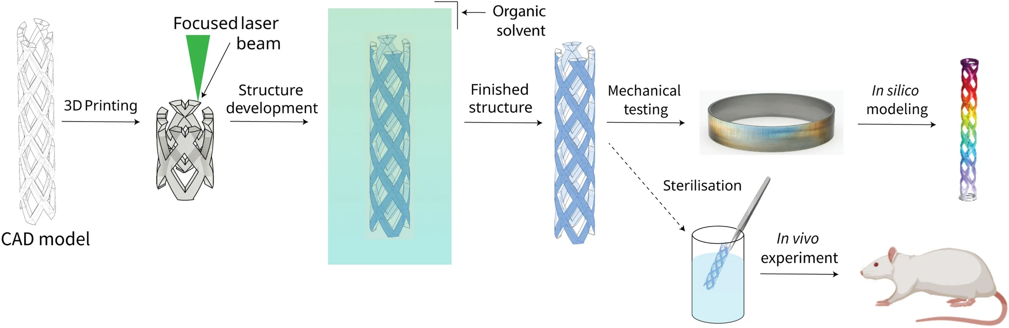
The schematics present the study workflow. First, CAD models of stents were generated. The printing process followed, with the goal of preparing structures for mechanical and biological testing. Additionally, cylinder samples were prepared from the same material as the stents. Mechanical testing was performed in two steps – experimental material characterization and in silico modeling. Finally, stents were validated using rats in vivo.
The World Health Organization (WHO) says that one of the leading causes of death globally are cardiovascular diseases (CVDs), including coronary artery disease (CAD), where the blood vessels narrow and become hard. A collaborative team from Lithuanian University of Health Sciences, Vilnius Gediminas Technical University, and Vital3D Technologies are using two-photon polymerization, or 2PP, 3D printing to fabricate stents to treat the narrowing of blood vessels. 3D printing is able to make complex architectural structures out of a variety of materials, making it attractive for producing medical devices like stents. But the technology is not without its limitations, such as some methods being unable to reach sub-μm levels of surface roughness, which medically viable stents need. So the team turned to 2PP, which is based on “nonlinear interaction between femtosecond laser light and photo-active resin” and has “unlimited 3D geometry potential.” They successfully used 2PP to 3D print stents for blood vessels as small as just 5 mm tall and 0.7 or 0.9 mm in diameter, and 3D struts as thin as 50 μm.
🏷️ p3d_feed“Several novel approaches were introduced to accommodate the printing of such a structure like voxel elongation and printing in stereolithography-like vat-sample holder configuration. Furthermore, the produced stents were tested mechanically proving their mechanical resilience to most common types of mechanical deformations. Experimental results were also compared to mathematical modeling, showing excellent agreement, hinting at the possibility of designing and testing complex micro-stent geometries before fabrication in silico. Finally, biocompatibility experiments were performed, in which rats survived the 7-day incubation period and showed no significant biocompatibility issues,” the researchers wrote in their study.