Stratasys’ RadioMatrix Could Mark the Beginning of the End for Cadavers in Imaging Training
⚓ p3d 📅 2025-12-03 👤 surdeus 👁️ 25

Stratasys has expanded its radiopaque 3D printing material, RadioMatrix, to full commercial availability in the United States. The move could change medical imaging training by giving hospitals and researchers access to lifelike, repeatable models that show up on X-rays and CT scans like real human tissue.
RadioMatrix is a photopolymer designed for Stratasys’ PolyJet printers, specifically the J750 and J850 Digital Anatomy systems. These machines already print soft tissue, bone, and vascular structures for surgical planning and education. Now, with RadioMatrix, they can also produce models that behave like real anatomy under medical imaging. Basically, the same printer used to mimic the feel of an organ can now mimic how that organ appears in a scan.
The key feature of RadioMatrix is control. Users can “tune” the radiopacity of the printed parts to match bone, grey matter, fat, veins, or any structure a radiologist expects to see. That means a hospital can print an entire organ with different regions matching the exact densities found in real patients.
To understand why this matters, we need to look into the Hounsfield unit (HU). CT scanners measure how dense a tissue is using this scale. According to experts, bone appears bright because it has a high HU value, while fat appears darker because it has a lower one. A material that looks realistic on a scan needs to fall within the correct HU range.
In fact, early work between Stratasys and Siemens Healthineers shows that RadioMatrix can match these values with incredible precision. According to the company, in some tests, deviations were as low as a single HU, which means the scanner can hardly tell the difference between a printed model and actual human tissue. This level of accuracy is rare for any “synthetic phantom” and is what gives RadioMatrix its true impact.
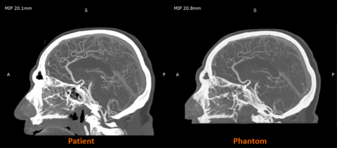
Patient versus Phantom imaging. Image courtesy of Stratasys.
Training Radiologists Without Cadavers
For decades, radiology education has relied heavily on cadavers and so-called “basic phantom blocks” made from plastics or gels. Still, both come with limits. Cadavers have a realism but are too expensive, rare, and even raise some ethical questions. Traditional phantoms are easier to access but don’t behave like human tissue. They also can’t be customized for specific pathology or printed in batches for consistent training.
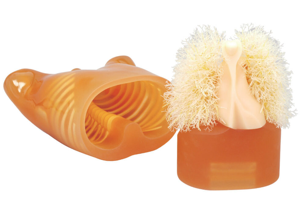
Multipurpose Chest Phantom N1 Lungman. Image courtesy of Kyoto Kagaku.
What’s more, ready-made phantoms and torso models for imaging training can cost thousands of dollars. A static adult male head phantom from Anatomy Warehouse costs $9,552, and an adult human torso phantom for X-ray, CT, and MRI training from GT Simulators costs $18,900. Specialized torso systems with more moving parts or higher complexity can cost $45,000 or more from brands like Kyoto Kagaku. Meanwhile, a traditional cadaver in the U.S. typically runs between $2,000 and $5,000, not including the extra costs of storage, preservation, lab space, and disposal. All of this helps explain why reusable, 3D printed radiopaque models such as RadioMatrix may attract interest from programs that already have 3D printing equipment.
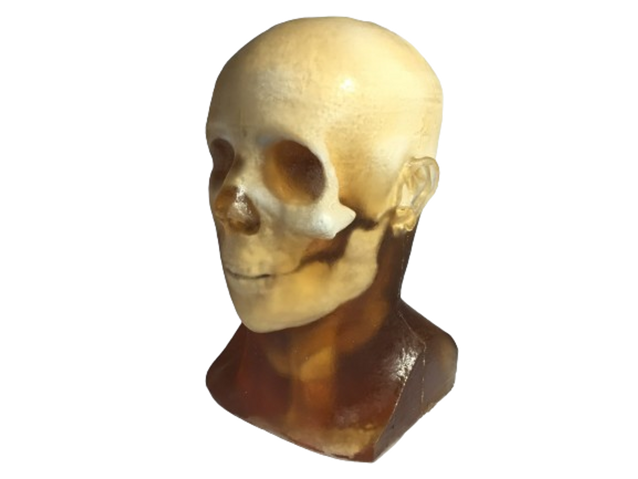
Static adult male head phantom model. Image courtesy of Anatomy Warehouse.
RadioMatrix is different. For example, a radiology department can print the same artery, tumor, or vessel structure many times. Students can practice with models that share the same target area, challenge, and path. Researchers can run controlled tests without waiting for donor material or relying on generic phantoms that don’t match what the clinical reality is all about.
The material is already being used in the UK at places like Beaumont Hospital and CPI. There, 3D printed cerebral angiography models are helping train doctors in imaging-guided procedures. These early tests show how printed radiopaque anatomy can make training more consistent and easier to repeat, especially for procedures that demand navigating tiny vessels.
In the U.S., there are many more medical schools and imaging centers than cadaver labs. Many smaller programs only get a few hours with shared donor bodies, or they rely on old training phantoms that don’t look realistic in modern CT scans. RadioMatrix gives these institutions an alternative.
The potential impact of the material extends to device testing as well. Companies testing new catheters, implants, or imaging tools often wait weeks for cadaver time, and the anatomy they get can vary from case to case. With RadioMatrix, they can print the exact anatomy they need and run tests as often as they want. In the end, that means they can have faster development cycles and fewer delays while waiting for donor material.
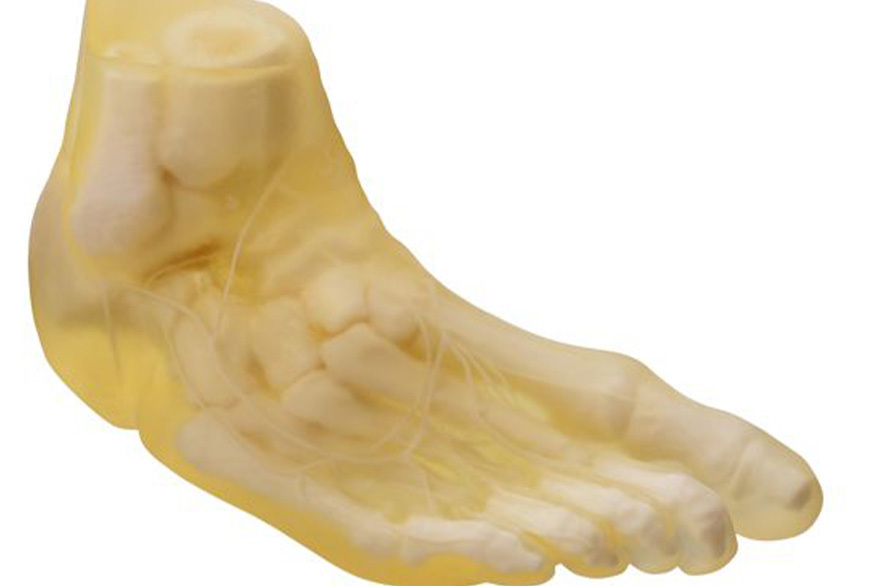
RadioMatrix material applications. Image courtesy of Stratasys.
Stratasys says the goal is not only to improve education, but also to strengthen how imaging algorithms are validated. By combining Digital Anatomy printing with accurate radiopacity, researchers can create realistic phantom datasets for AI and imaging software, datasets that look identical, scan after scan. This is nearly impossible to achieve with cadavers or traditional phantoms.
“Providing full availability of RadioMatrix in the U.S. is a major step in providing cutting-edge imaging education and training,” said Erez Ben Zvi, Vice President of Healthcare at Stratasys. “By giving radiologists and device manufacturers the ability to print ultra-realistic, customized radiographically accurate models, we’re helping replace traditional phantom solutions and reliance on cadavers with customizable, repeatable, and scalable alternatives.”
Now that RadioMatrix is available in the U.S., hospitals and researchers can choose to incorporate it into their training and testing workflows. What’s more, the impact will become clearer as more institutions begin to use it.
🏷️ p3d_feed